Optical Coherence Tomography Assessment of Apparent Foveal Swelling in Patients with Foveal Sparing Secondary to Geographic Atrophy. 26/03/2013

OBJECTIVE:
To determine whether foveal swelling exists in patients with foveal sparing and geographic atrophy (GA) secondary to dry age-related macular degeneration (AMD) and to establish the contribution of different foveal layers to this condition by use of spectral-domain optical coherence tomography (SD-OCT).
DESIGN:
Prospective comparative case series.
PARTICIPANTS:
We assessed patients from a longitudinal study with foveal sparing and GA secondary to AMD. Of an initial sample of 108 patients, 13 eyes of 10 patients complied with the inclusion criteria to study eyes in which apparent swelling would not be questionable. We used a control group of 13 healthy patients to compare the outcome measurements.
METHODS:
We acquired high-resolution SD-OCT horizontal and oblique B-scans centered at the umbo. Two retinal specialists (J.M., F.T.) independently classified the SD-OCT images.
MAIN OUTCOME MEASURES:
Difference in foveal center thickness, apparent outer nuclear layer (ONL) thickness, ONL thickness without Henle's fiber layer (HFL), sub-ONL thickness, and retinal thickness at 1000 μm and 3500 μm from the foveal center.
RESULTS:
The thickness at the foveal center was similar between patients with apparent foveal swelling (cases) and controls without AMD (226 vs. 227 μm; P = 0.56), but the apparent ONL was thicker in cases than in controls (125 vs. 114 μm; P = 0.02). However, when HFL was excluded from the measurements, there was little difference in the results (74 vs. 73 μm; P = 0.82).
CONCLUSIONS:
We found neither foveal nor ONL swelling in this study. We observed HFL thickening in foveal sparing secondary to GA, which might be related to swelling of the axons of the photoreceptors, or Müller's cells. We also observed thinning of the retina below the external limiting membrane. The clinical significance of these findings should be addressed by longitudinal studies and may have specific therapeutic implications.
FINANCIAL DISCLOSURE:
Proprietary or commercial disclosure may be found after the references.
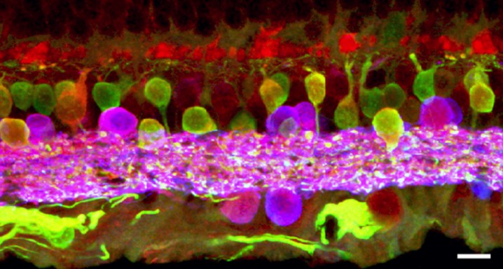
Image source: Gema Martínez-Navarrete
DMAE seca o atróficaDMAE exudativa o húmedaAngiografía fluoresceínicaAutofluorescenciaRetinografíaTomografía de coherencia ópticaMicroperimetría


