LITE: Development of Advanced Laser Imaging Techniques for the anterior and posterior Eye 30/01/2015

Description
Development of Advanced Laser Imaging Techniques for the Anterior and Posterior Eye (LITE) is a project involving the creation of a hybrid system that will allow for new eye imaging techniques to treat pathologies of the retina and cornea.
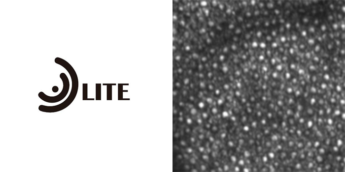
These new techniques will enable high-resolution images of the retina’s photoreceptors to be obtained through the latest adaptive optical technique, together with the collagen fibres of the cornea.
Heading image: Courtesy of Prof. Jacque Duncan, Prof. Austin Roorda and Dr. David Merino.
Key inclusion criteria
Age-related macular degeneration (AMD) in its atrophic form, pigmentary retinosis and Stargardt’s Disease.
Project Objectives
The new system will provide a diagnosis that is better tailored to the cellular level, enabling improved knowledge of retinal diseases and offering new, more precise effectiveness assessment parameters. This responds to the new emerging experimental therapies, such as regenerative medicine treatments with stem cell implantation, for the degenerative retinal diseases currently lacking effective treatment and which are associated with severe vision loss like age-related macular degeneration (AMD) in its atrophic form, pigmentary retinosis and Stargardt’s Disease.
Moreover, the new system of advanced imaging techniques will allow for treatment of the anterior and posterior eye segments at the same time, thereby reducing the costs of equipment.
Publications
-
Merino, D., & Loza-Alvarez, P. (2016). Adaptive optics scanning laser ophthalmoscope imaging: technology update. Clinical ophthalmology (Auckland, NZ), 10, 743.
-
Rossi, F., Canovetti, A., Malandrini, A., Lenzetti, I., Pini, R., & Menabuoni, L. (2015). An “All-laser” Endothelial Transplant. JoVE (Journal of Visualized Experiments), (101), e52939-e52939.
-
Psilodimitrakopoulos, S., Loza-Alvarez, P., & Artigas, D. (2014). Fast monitoring of in-vivo conformational changes in myosin using single scan polarization-SHG microscopy. Biomedical optics express, 5(12), 4362-4373.
-
Cicchi, R., Baria, E., Matthäus, C., Lange, M., Lattermann, A., Brehm, B. R., ... & Pavone, F. S. (2015). Non‐linear imaging and characterization of atherosclerotic arterial tissue using combined SHG and FLIM microscopy. Journal of biophotonics, 8(4), 347-356.
-
Cicchi, R., Rossi, F., Alfieri, D., Bacci, S., Tatini, F., De Siena, G., ... & Pavone, F. S. (2016). Observation of an improved healing process in superficial skin wounds after irradiation with a blue‐LED haemostatic device. Journal of biophotonics.
-
Mercatelli, R., Ratto, F., Rossi, F., Tatini, F., Menabuoni, L., Malandrini, A., ... & Cicchi, R. (2016). Three‐dimensional mapping of the orientation of collagen corneal lamellae in healthy and keratoconic human corneas using SHG microscopy. Journal of Biophotonics.
Funded by the BiophotonicsPlus. Programa ERA-NET . 7º PM. European Comission. ACC1Ó, Agència per a la competitivitat de l'empresa, Departament d'Empresa i Ocupació de la Generalitat de Catalunya.
Author
Dr. Jordi Monés, M.D., Ph.D.
COMB Medical license number: 22.838
Director
Doctor of Medicine and Surgery
Specialist in Ophthalmology
Specialist in Retina, Macula and Vitreorretinal


